

Canine skin disease is the most common reason dogs present to veterinary practices. A significant proportion of those cases — atopic dermatitis, seborrhea, zinc-responsive dermatosis, post-glucocorticoid skin changes — have a nutritional or barrier-lipid component amenable to targeted supplementation. Understanding the mechanism behind each nutrient clarifies both what to expect and the realistic timeline for improvement.
The skin barrier: a lipid-dependent structure
The epidermal barrier function depends on a precise mixture of lipids secreted by keratinocytes into the intercellular space of the stratum corneum: approximately 50% ceramides, 25% cholesterol, and 15% free fatty acids, arranged in lamellar bilayers. This lipid matrix controls transepidermal water loss (TEWL) and prevents penetration of allergens, irritants, and pathogens. Disruption of any component — ceramide depletion in atopic dermatitis, fatty acid imbalance from poor diet, free radical damage to membrane lipids — increases TEWL, permits allergen penetration, and increases inflammatory sensitization.
Omega-3 fatty acids, zinc, biotin, and vitamin E each address different components of this barrier system. Their combined effect is greater than any single agent alone.
Omega-3 EPA+DHA: the primary skin supplement
EPA and DHA exert their dermatological effects through several mechanisms. EPA competes with arachidonic acid for cyclooxygenase and lipoxygenase enzymes, reducing production of pro-inflammatory prostaglandins (PGE2) and leukotrienes (LTB4) — the inflammatory mediators that drive pruritus, erythema, and mast cell degranulation in atopic skin. DHA incorporates into keratinocyte and sebocyte membrane phospholipids, improving membrane fluidity and structural integrity. At the barrier level, omega-3 supplementation increases ceramide synthesis and reduces TEWL in atopic dogs — a direct barrier restoration effect documented in veterinary dermatology studies.
Therapeutic dose matters: studies showing clinical benefit in atopic dogs use 40–180 mg EPA+DHA per kilogram bodyweight per day. Products providing 5–10 mg per pound are sub-therapeutic for dermatological purposes. The EPA:DHA ratio is less critical than total EPA+DHA dose.
Zinc: essential for keratinocyte function
Zinc is a cofactor for over 300 enzymes, including those involved in keratinocyte proliferation, differentiation, and cornification. Zinc deficiency — or zinc malabsorption — produces a specific clinical syndrome: zinc-responsive dermatosis. In dogs, two presentations occur:
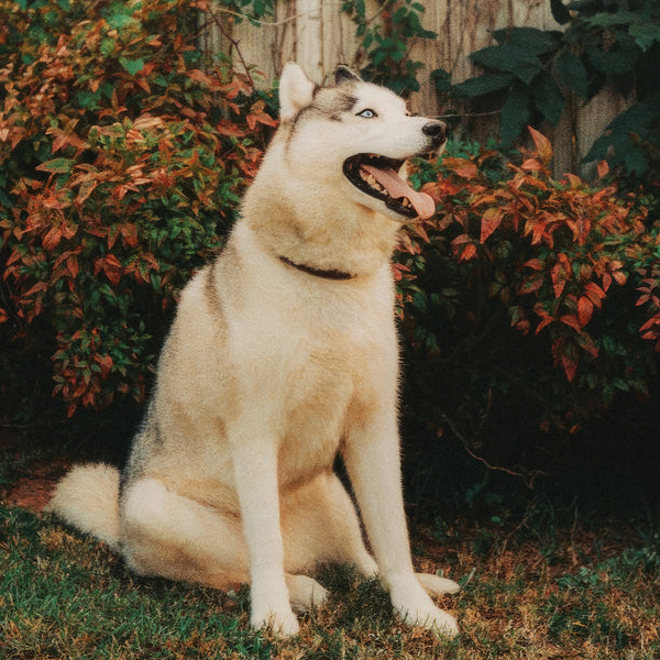
Syndrome I: Primarily in Siberian Huskies and Alaskan Malamutes, caused by a genetically determined impairment in intestinal zinc absorption. These dogs develop crusting and scaling around the face (muzzle, periocular, ear margins) despite adequate dietary zinc. The lesions are characteristic and respond dramatically to zinc supplementation — often within 4–6 weeks. This is a genuine clinical entity with a known mechanism, not a theoretical supplement benefit.
Syndrome II: Occurs in rapidly growing large breed puppies fed zinc-deficient or high-phytate diets (phytate chelates zinc and reduces absorption). Scaling and crusting in a generalized distribution; resolves with diet correction and zinc supplementation.
Beyond zinc-responsive dermatosis, zinc supports wound healing, collagen synthesis, and keratinocyte proliferation generally — relevant in any dog with compromised skin integrity. Zinc methionine and zinc gluconate have better bioavailability than zinc oxide.
Biotin: cofactor for fatty acid synthesis
Biotin (vitamin B7) is a cofactor for acetyl-CoA carboxylase, the rate-limiting enzyme in de novo fatty acid synthesis, and for other carboxylase enzymes involved in lipid and amino acid metabolism. In the context of skin health, biotin supports the synthesis of long-chain fatty acids that are incorporated into ceramides and membrane phospholipids — directly relevant to barrier function. Biotin deficiency produces a dull, dry coat with scaling and brittleness. Commercial diets generally provide adequate biotin, but dogs with malabsorptive conditions, chronic antibiotic use (which disrupts biotin-producing gut bacteria), or high raw egg white intake (avidin blocks biotin absorption) are at risk.
Even without clinical deficiency, biotin supplementation at 1,000–5,000 mcg/day supports ceramide synthesis substrate availability and is appropriate as part of a barrier-restoration protocol.
Vitamin E: membrane antioxidant for keratinocytes
Vitamin E (alpha-tocopherol) is the primary lipid-soluble antioxidant in cell membranes, where it quenches free radicals and prevents lipid peroxidation. Keratinocytes and sebocytes are exposed to UV radiation, inflammatory oxidative stress, and polyunsaturated fatty acid peroxidation — all of which deplete membrane vitamin E. Dogs on high omega-3 diets or supplementation have increased PUFA content in membranes and therefore increased antioxidant demand; vitamin E supplementation is specifically indicated alongside omega-3 to prevent peroxidative membrane damage. The synergy between omega-3 and vitamin E is well-documented: EPA+DHA incorporation into membranes without adequate vitamin E increases peroxidative vulnerability.
Condition-by-condition supplement approach
| Condition | Primary supplement approach | Timeline for improvement |
|---|---|---|
| Atopic dermatitis | Omega-3 at therapeutic dose + quercetin; biotin + zinc for barrier support | 4–8 weeks for itch reduction; 8–12 weeks for full barrier effect |
| Primary seborrhea | Omega-3 + zinc + biotin; vitamin E; consider evening primrose oil for GLA | 6–10 weeks for scale reduction |
| Zinc-responsive dermatosis | Zinc supplementation (methionine or gluconate form); correct underlying absorption issue | 4–6 weeks for dramatic improvement |
| Post-glucocorticoid skin thinning | Omega-3 (partially restores barrier lipids); biotin + zinc for keratinocyte support | 8–16 weeks; improvement limited by degree of chronic steroid damage |
| Demodex | Antiparasitic treatment is primary; omega-3 for immune support as adjunct only | Supplements do not treat active demodicosis |
| Bacterial pyoderma | Antibiotics are primary; omega-3 + zinc support healing as adjuncts | Supplements do not replace antibiotic treatment |
| Dermatophytosis (ringworm) | Antifungal treatment is primary; no supplement role in eliminating infection | Supplements do not treat ringworm |
Conditions where supplementation is insufficient alone
Demodicosis, bacterial pyoderma, and dermatophytosis all have infectious or parasitic etiologies that require primary treatment — antiparasitic, antibiotic, or antifungal respectively. Supplements support healing and immune function as adjuncts but cannot resolve the primary pathology. Owners who attempt to manage active secondary infections with supplements alone risk allowing the condition to become more deeply established and difficult to treat.
Related: coat supplement guide · allergy supplement · omega-3 for dogs · biotin, zinc, and vitamin E guide · gut-skin connection.